2.15B - How the eye works
Follow light through the eye, explain how rods and cones produce sensory information, and distinguish the iris response to light from lens accommodation. This Biology-only lesson applies to both tiers.
2.15B — From light to an electrical signal
The eye is a sense organ. Its stimulus is visible light, and its receptor cells are in the retina at the back of the eye. The structures at the front of the eye first control and focus the light; the receptor cells then convert the light stimulus into electrical impulses.
Sensory receptor
A sensory receptor is a cell or group of cells that detects a stimulus and converts it into an electrical impulse.
Light follows this route:
cornea -> pupil -> lens -> retina -> electrical impulses in the optic nerve
The direction matters. The retina does not focus light, and the cornea does not detect it. The diagram is a simplified route: both the cornea and lens refract light, while the arrow along the optic nerve represents electrical impulses, not travelling light.
| Structure | Feature and function | Contribution to vision |
|---|---|---|
| Cornea | A transparent, curved surface at the front of the eye. It refracts light, which means that it bends the rays as they enter. | Provides much of the initial focusing needed to direct light towards the retina. |
| Pupil | An opening in the centre of the iris. | Allows light to enter the eye. The pupil is a gap, not a muscle. |
| Iris | A ring containing muscles around the pupil. | Changes pupil diameter and therefore controls how much light enters. |
| Lens | A transparent, flexible structure behind the iris. It refracts light and can change shape. | Fine-tunes the focus so rays from the viewed object meet on the retina. |
| Retina | Light-sensitive tissue lining the back of the eye. It contains rod and cone receptor cells. | Converts the light stimulus into electrical impulses. |
| Optic nerve | A bundle of nerve fibres leaving the retina. | Carries electrical impulses from the retina towards the brain. |
[DIAGRAM: asset_name: 2.15B-2.17B - The eye and vision correction - diagram 01; asset_slug: edexcel-gcse-biology-2-15b-2-17b-eye-sensory-route; recommended_method: image_gen; description: Simple monochrome textbook cross-section of one human eye showing incoming light travelling in the correct direction through cornea, pupil and lens to the retina, then an arrow labelled electrical impulses leaving along the optic nerve. Label only cornea, iris, pupil, lens, retina, rod cells, cone cells and optic nerve; include a small retinal inset that locates rods and cones without histological detail; make every leader line unambiguous; do not add off-scope structures, photorealism, colour coding, a clinical scan or an observed specimen.]
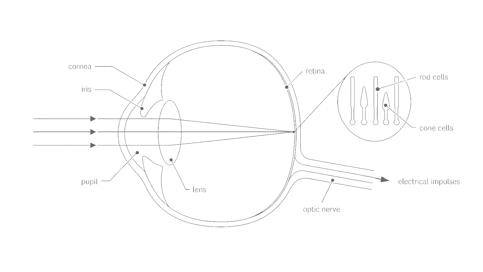
Rod and cone cells are both photoreceptors, but they are suited to different light conditions:
| Receptor cell | Sensitivity and role |
|---|---|
| Rod cell | Very sensitive to light, so it supports vision in dim conditions. Rods detect light intensity but do not distinguish colours. |
| Cone cell | Less sensitive than a rod, so it works best in brighter light. Different cone types respond most strongly to different wavelength ranges, allowing colour vision and fine detail. |
When light stimulates either type, the retina produces electrical signals. These are passed towards the brain, where the visual information is processed.
2.15B — Controlling light and focus
Two adjustments keep the light reaching the retina useful. The iris controls the amount of light, while the lens adjusts the focus for objects at different distances. These are different jobs: the iris does not focus the image, and the lens does not change pupil size.
The iris and pupil
The iris contains circular and radial muscles. Their opposing actions alter the diameter of the pupil:
| Light condition | Iris muscle action | Pupil response | Result |
|---|---|---|---|
| Bright light | Circular muscles contract and radial muscles relax. | The pupil constricts: it becomes narrower. | Less light reaches the retina, reducing excessive stimulation of the receptor cells. |
| Dim light | Radial muscles contract and circular muscles relax. | The pupil dilates: it becomes wider. | More light reaches the retina, helping sensitive rod cells respond. |
The pupil itself does not contract. It is the opening whose size is changed by muscles in the iris.
The lens and accommodation
Accommodation
Accommodation is the change in lens shape that keeps light from objects at different distances focused on the retina.
Light from a nearby object needs more refraction than light from a distant object. The lens changes shape through the ciliary muscles and suspensory ligaments:
| Viewed object | Ciliary muscles | Suspensory ligaments | Lens | Optical result |
|---|---|---|---|---|
| Near | Contract | Become slack | Becomes thicker and more convex | Refracts light more strongly so it focuses on the retina. |
| Distant | Relax | Become taut | Becomes thinner and less convex | Refracts light less strongly so it focuses on the retina. |
The counterintuitive step is worth noticing: contraction of the ciliary muscles makes the suspensory ligaments less tense. This releases their pull on the elastic lens, allowing it to become thicker.
Iris muscles alter how much light enters; ciliary muscles alter how strongly the lens refracts that light.