2.27-2.28 - Alimentary canal and peristalsis
Food does not just sit inside the body while it is digested. It passes through a muscular tube called the alimentary canal, where different regions carry out different jobs. This lesson follows the route food takes and then explains the muscle action that keeps it moving.
The alimentary canal as a route
The alimentary canal is the long tube that food passes through during digestion. In this cluster, the required route is:
mouth -> oesophagus -> stomach -> small intestine -> large intestine -> rectum
The pancreas is also required, but it is not a section of the tube that food passes through. It is an associated organ that releases digestive substances into the small intestine.
Alimentary Canal
The alimentary canal is the muscular tube through which food passes as it is digested and prepared for removal from the body.
[DIAGRAM: asset_name: Alimentary canal and peristalsis - diagram 01; asset_slug: b12_alimentary_canal_and_peristalsis__diagram_01; recommended_method: deterministic_drawing; description: Monochrome schematic of the human alimentary canal labelled mouth, oesophagus, stomach, pancreas, duodenum, ileum, colon and rectum, with arrows showing movement from mouth to rectum. The pancreas is shown beside the duodenum as an associated organ, not part of the continuous food pathway.]
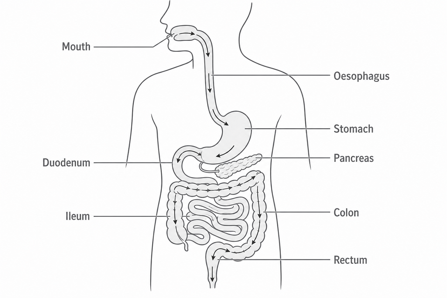
| Structure | Position in the route | Main function in this lesson |
|---|---|---|
| Mouth | First part | Takes in food and chews it into smaller pieces. |
| Oesophagus | Between mouth and stomach | Moves swallowed food to the stomach. |
| Stomach | After the oesophagus | Stores, churns and mixes food. |
| Duodenum | First part of the small intestine | Receives food from the stomach and digestive substances from the pancreas. |
| Ileum | Later part of the small intestine | Main region where digested food molecules are absorbed. |
| Colon | Main part of the large intestine | Absorbs water from the remaining material. |
| Rectum | End of the large intestine | Stores faeces before egestion. |
| Pancreas | Beside the duodenum | Produces pancreatic juice containing digestive enzymes. |
Mouth, oesophagus and stomach
The mouth is where food enters the alimentary canal. Chewing breaks food into smaller pieces, making it easier to swallow and increasing the surface area that digestive enzymes can act on later. Saliva also moistens food so that it can be formed into a soft bolus.
The oesophagus is a muscular tube that connects the mouth to the stomach. Its job is transport, not digestion. Swallowed food is moved along it by peristalsis, which is the process explained in the final part.
The stomach is a muscular bag. It stores food for a time, churns it, and mixes it with acidic digestive juice. At this level, the key function is that the stomach physically mixes food and begins turning it into a softer mixture that can enter the small intestine.
The mouth prepares food for swallowing, the oesophagus transports it, and the stomach stores and churns it.
Duodenum, ileum and pancreas
The small intestine has two named parts in this specification point: the duodenum and the ileum.
The duodenum is the first part of the small intestine. It receives partly digested food from the stomach. It also receives pancreatic juice from the pancreas, so it is a key place where digestion continues.
The ileum is the later part of the small intestine. Its main function is absorption: small, soluble products of digestion pass from the ileum into the blood. The detailed adaptations of the ileum are covered in the next digestion cluster, so here the important point is the function of the region.
The pancreas is not part of the alimentary canal route because food does not pass through it. Its required function here is to make pancreatic juice, which contains digestive enzymes, and release it into the duodenum.
Colon and rectum
The large intestine comes after the small intestine. In this specification point, the named parts are the colon and the rectum.
The colon absorbs water from the remaining undigested material. This changes the contents from a more liquid mixture into more solid faeces. This is why the colon is linked with water absorption rather than with absorbing most digested food molecules.
The rectum is the final storage region. It stores faeces before they leave the body. In exam answers, keep the functions separate: the colon absorbs water; the rectum stores faeces.
How peristalsis moves food
Peristalsis is the wave-like movement of the gut wall that pushes food through the alimentary canal. It happens because muscles in the wall of the gut contract and relax in a coordinated sequence.
Peristalsis
Peristalsis is the movement of food through the gut by waves of muscle contraction and relaxation in the gut wall.
[DIAGRAM: asset_name: Alimentary canal and peristalsis - diagram 02; asset_slug: b12_alimentary_canal_and_peristalsis__diagram_02; recommended_method: deterministic_drawing; description: Monochrome three-panel sequence showing a food bolus inside a gut tube. Circular muscle behind the bolus contracts to squeeze it forward, muscle ahead relaxes, and arrows show the direction of movement.]
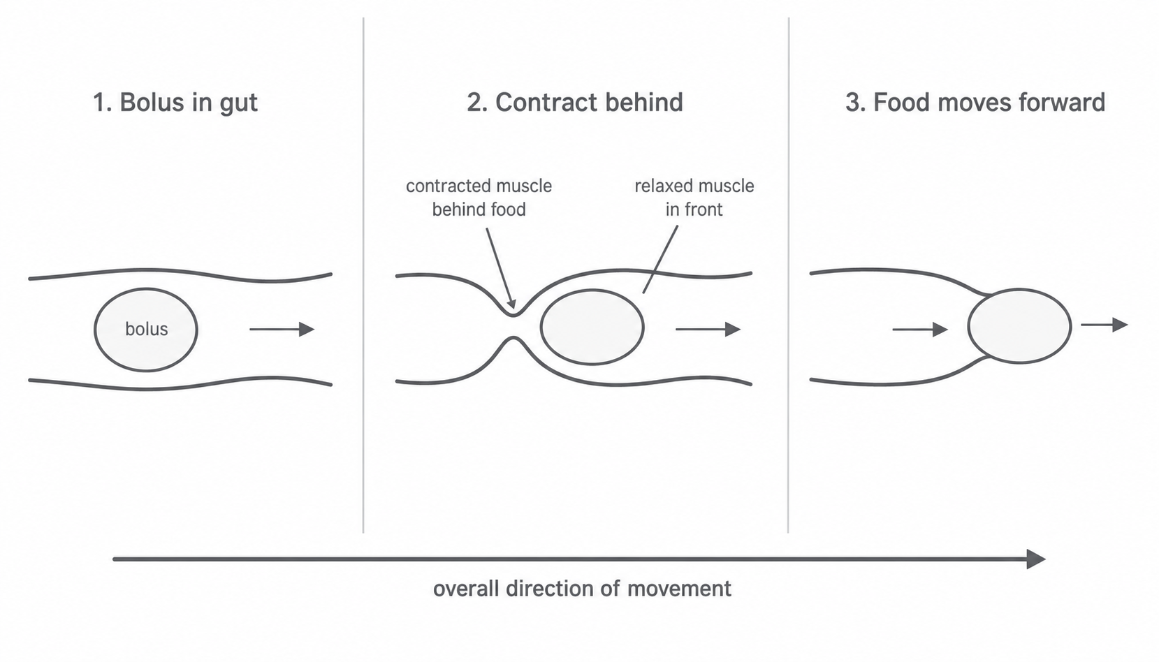
Think of a bolus of food inside the gut:
- Muscle behind the bolus contracts.
- This narrows the gut behind the food and squeezes it forward.
- Muscle in front of the bolus relaxes.
- The relaxed region lets the food move into the next section.
Peristalsis is important because food needs to move through the gut even when gravity is not helping. It is also not the same as digestion: digestion breaks large food molecules into smaller soluble molecules, while peristalsis moves and mixes food along the canal.
Common exam traps are to say that gravity moves food through the gut, to describe peristalsis as food "falling", or to put the pancreas in the route food follows. The mark-winning idea is coordinated contraction and relaxation of gut muscles.