4.2.2 - Neurons and synaptic transmission
Neural communication depends on specialised cells carrying impulses in the correct direction and passing signals across tiny gaps. You will compare sensory, relay and motor neurons, linking dendrons, cell bodies, axons and terminals to their roles in a response pathway. The lesson then follows synaptic transmission from vesicle release to receptor binding, showing how neurotransmitters can excite or inhibit the postsynaptic neuron.
Neurons as communication cells
Neurons receive, carry and pass on information using specialised structures.
AO1: the basic job of a neuron
The nervous system works because specialised cells can carry information quickly from one place to another. These cells are neurons. A neuron can receive information, pass an electrical impulse along its length, and communicate with another cell at the end.
Neuron
A neuron is a specialised nerve cell that transmits information through the nervous system using electrical impulses within the cell and chemical communication between cells.
Most AQA answers need the same core structure:
- Dendrites receive signals from other neurons or from sensory receptors.
- The cell body, or soma, contains the nucleus and keeps the neuron alive.
- The axon carries the electrical impulse away from the cell body.
- The myelin sheath is a fatty insulating layer around many axons that helps impulses travel faster.
- Nodes of Ranvier are gaps in the myelin sheath. In myelinated neurons, the impulse effectively jumps from node to node.
- Axon terminals or terminal boutons are the endings of the axon. They form synapses with the next cell.
Nerve impulse
A nerve impulse is an electrical signal, often called an action potential, that travels along a neuron.
AO2: why structure matters
Imagine touching a sharp pin. The receptor in the skin detects the stimulus. A neuron must carry that information towards the spinal cord or brain, another neuron may process or relay it inside the central nervous system, and another neuron may carry a command to a muscle. The structure of each neuron helps it do its particular job in that pathway.
The big exam habit is to link structure to function. Do not only name parts. Say what they do. For example, "the axon carries the impulse away from the cell body" is stronger than simply writing "the neuron has an axon".
[DIAGRAM: asset_name: Sensory relay motor neuron comparison; asset_slug: 4_2_2_02_the_structure_and_function_of_sensory_relay_and_motor_neurons__diagram_01; recommended_method: image_gen; description: Comparison of sensory, relay and motor neurons in three columns, with impulse direction and labels for receptor to CNS, within CNS, CNS to effector, axon, cell body and dendrites.]
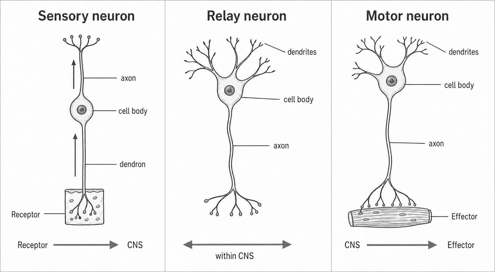
The sensory drawing is a simplified shape, not a universal template. Its upward arrows show receptor-to-CNS transmission; the relay panel’s horizontal double arrow denotes its location within the CNS, not two-way transmission through one chemical synapse. Typical skin sensory neurons have the cell body off the main pathway, as described below.
Sensory, relay and motor neurons
The three functional types differ in the direction and destination of the information they carry.
AO1: three functional types
Sensory neuron
A sensory neuron carries impulses from sensory receptors towards the central nervous system.
Sensory neurons are sometimes called afferent neurons because they carry information arriving at the CNS. Their job is input. They respond to physical or chemical stimuli such as heat, pressure, light, sound or chemicals involved in taste and smell. Many sensory neurons have a cell body positioned off to the side of the main axon, which is why their shape can look different from the textbook "tree" shape.
Relay neuron
A relay neuron is a neuron inside the central nervous system that connects sensory neurons with other neurons, including motor neurons.
Relay neurons are also called interneurons in many biology sources. AQA uses "relay neuron", so use that term in exam answers. Their job is processing and connection inside the brain or spinal cord. They often have many dendrites because they may receive information from several neurons and pass it on within a neural circuit.
Motor neuron
A motor neuron carries impulses from the central nervous system to effectors such as muscles or glands.
Motor neurons are sometimes called efferent neurons because they carry commands exiting the CNS. Their job is output. A motor neuron may have a cell body at one end, a long axon, and terminals that connect to an effector. If the effector is a muscle, the result may be contraction.
AO2: applying the pathway
In a simple withdrawal reflex, Aisha touches a very hot baking tray:
- Receptors in her skin detect heat and tissue damage.
- A sensory neuron carries an impulse from the receptor towards the spinal cord.
- A relay neuron in the spinal cord passes the message across the CNS pathway.
- A motor neuron carries an impulse from the spinal cord to muscles in her arm.
- The muscles contract, pulling her hand away.
This is useful AO2 because the same three-neuron logic can be applied to many scenarios: pain, pressure, movement, glands, or any stimulus-response example.
How structure supports function
The shape and connections of a neuron support its role in a communication pathway.
AO1: comparing the shapes
All three neuron types use the same basic principles, but their structure is adapted to their role.
Sensory neurons need to carry information from receptors over potentially long distances towards the CNS. In the common textbook sensory neuron, a long dendron carries the impulse from the receptor towards the cell body, which lies to one side; a shorter axon carries it towards the CNS. Real sensory neurons vary in shape, so these relative lengths describe a typical example rather than every sensory neuron.
Relay neurons are usually found in the brain and spinal cord. They tend to be shorter and highly branched, which suits their role in connecting many neurons inside the CNS. The many dendrites and synaptic connections allow a relay neuron to integrate information before passing it on.
Motor neurons carry commands away from the CNS to effectors. A long axon lets a command travel from the spinal cord towards a muscle or gland. Branching terminals allow one motor neuron to communicate with muscle fibres or other effector cells.
AO2: deciding from clues
If a scenario says a neuron carries an impulse from a receptor to the spinal cord, it is sensory. If it says the neuron is inside the brain or spinal cord and links other neurons, it is relay. If it says the neuron carries an impulse from the CNS to a muscle or gland, it is motor.
That is usually more reliable than trying to memorise one fixed drawing. Real neurons vary widely, so exam answers should prioritise the function and the direction of travel.
AO3: a useful but simplified classification
The sensory-relay-motor distinction is useful because it makes stimulus-response pathways clear. It is also a simplification. In the brain, neurons are much more diverse than three neat categories, and many are classified by neurotransmitter, connection pattern or electrical properties. For AQA, that complexity is background only. The exam target is the functional difference between the three named types.
Synaptic transmission
At a chemical synapse, neurotransmitter links the electrical activity of one neuron to a response in the next cell.
AO1: what happens at a synapse
Neurons do not usually touch end-to-end. The whole junction is a synapse; its tiny gap is the synaptic cleft. The signal travels electrically along the axon, but it crosses most synapses chemically.
Synapse
A synapse is the junction between a neuron and another cell, where information is passed across a tiny gap called the synaptic cleft.
Neurotransmitter
A neurotransmitter is a chemical messenger released by a presynaptic neuron that crosses the synaptic cleft and binds to receptors on the postsynaptic cell.
[DIAGRAM: asset_name: Synaptic transmission sequence; asset_slug: 4_2_2_02_the_structure_and_function_of_sensory_relay_and_motor_neurons__diagram_02; recommended_method: image_gen; description: Chemical synaptic transmission with presynaptic neuron, vesicles, neurotransmitter, synaptic cleft, receptors, postsynaptic neuron and the possible excitation or inhibition response.]
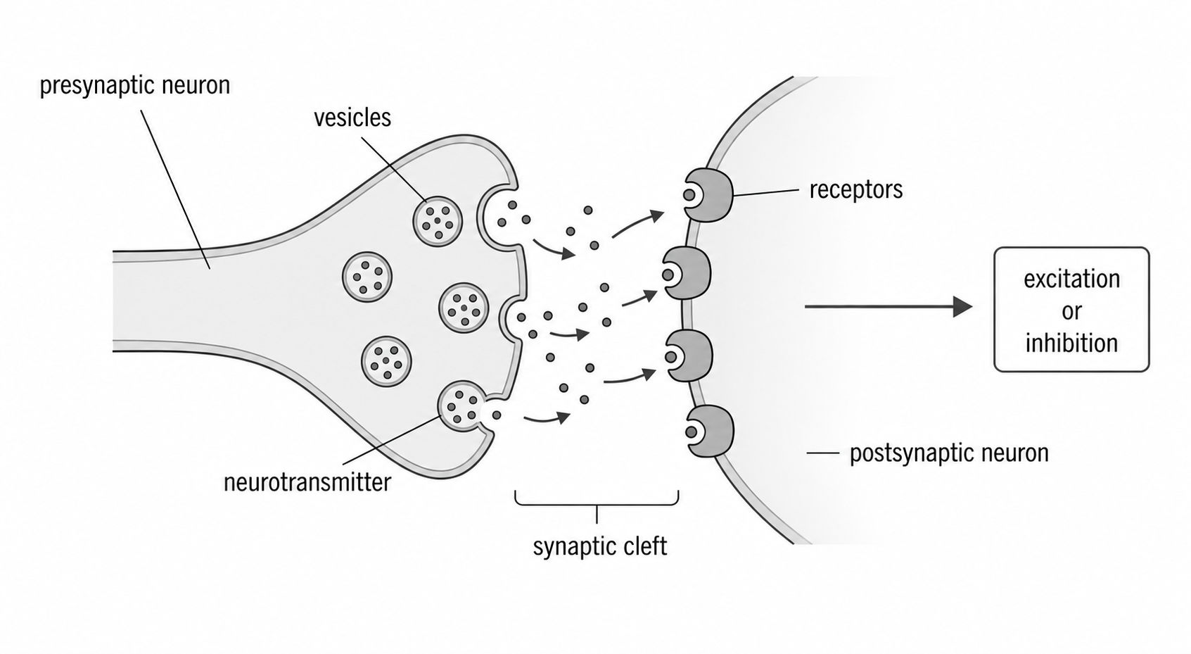
The process can be described in a clear sequence:
- An action potential reaches the axon terminal of the presynaptic neuron.
- Vesicles in the presynaptic terminal release neurotransmitter into the synaptic cleft.
- Neurotransmitter diffuses across the synaptic cleft.
- Neurotransmitter binds to specific receptor sites on the postsynaptic neuron.
- The postsynaptic neuron is affected. The effect may be excitatory or inhibitory.
- Neurotransmitter is removed from the synaptic cleft, for example by breakdown or reuptake, so the signal is brief and controlled.
Chemical transmission normally proceeds in one direction at this junction: transmitter is released from presynaptic vesicles and detected by receptors on the postsynaptic membrane. This is different from claiming that a whole brain circuit can never feed information back.
AO2: active pathway example
If the presynaptic neuron is part of a pain pathway, the chemical message at the synapse helps pass information to the next neuron in the spinal cord or brain. If the postsynaptic neuron reaches threshold, it may generate its own action potential and continue the pathway.
Excitation and inhibition
Postsynaptic signals change the likelihood of an action potential, rather than guaranteeing a response.
AO1: two possible postsynaptic effects
AQA specifically names excitation and inhibition, so these terms need to be secure.
Excitation
Excitation means a postsynaptic effect that increases the likelihood that the postsynaptic neuron will fire an action potential.
An excitatory effect usually involves depolarisation: the postsynaptic membrane becomes less negative and moves closer to the threshold needed to fire. An excitatory neurotransmitter does not guarantee that the next neuron fires. It increases the chance.
Inhibition
Inhibition means a postsynaptic effect that decreases the likelihood that the postsynaptic neuron will fire an action potential.
An inhibitory effect may involve hyperpolarisation: the postsynaptic membrane becomes more negative and moves further from threshold. This makes an action potential less likely.
The postsynaptic neuron often receives many inputs at once. Their combined effect is called summation; the strength and timing of the inputs matter, not just the number of excitatory and inhibitory synapses. Some are excitatory and some are inhibitory. Whether it fires depends on the balance of these inputs. This is why neural communication is not just an on/off chain. It is an integrated system of signals.
AO2: applying excitation and inhibition
Suppose a relay neuron in the spinal cord receives two excitatory messages and one inhibitory message at the same time. If the excitatory effects are strong enough overall, the relay neuron may fire and pass the message on. If inhibition dominates, it may not fire. In behaviour terms, this can affect whether a response is triggered, strengthened or held back.
AO3: why the detail matters
This biological explanation is powerful because it shows how behaviour can be linked to physical processes in the nervous system. It also shows why "a neurotransmitter causes behaviour" is too simple. Neurotransmitters act at particular receptors, within particular circuits, and their effects depend on the balance of excitation and inhibition.
That gives you a neat evaluation point for later biopsychology topics: neural explanations are scientific and precise, but they can become biologically reductionist if they ignore cognition, social context and wider psychological explanations.