3.2.3 - Transport Across Cell Membranes
Cells survive because their membranes are selective rather than sealed shut. In this lesson, we move from the fluid-mosaic structure of membranes to the main transport mechanisms, and then finish with the adaptations that make transport fast in specialised cells.
Part 1: Membrane Structure and Selective Permeability
All cell-surface membranes and organelle membranes share the same basic plan. This is described by the fluid-mosaic model.
Fluid-Mosaic Model
A model in which phospholipids form a fluid bilayer and proteins are scattered through it like a mosaic. Individual phospholipids can move sideways within the layer.
Phospholipids arrange themselves into a bilayer because their phosphate heads are hydrophilic and their fatty acid tails are hydrophobic. The heads face the watery environments on either side of the membrane, while the tails face inwards away from water.
This arrangement matters. The hydrophobic middle of the membrane allows small non-polar molecules such as oxygen and carbon dioxide to cross quite easily, but it makes life much harder for ions and large or polar molecules.
| Membrane component | Main role |
|---|---|
| Phospholipids | Form the bilayer and create a barrier to many water-soluble substances |
| Channel proteins | Form hydrophilic pores for particular ions or water molecules |
| Carrier proteins | Bind to a specific substance and change shape to move it across |
| Glycoproteins | Act in cell recognition and as receptors |
| Glycolipids | Help with recognition and membrane stability |
| Cholesterol | Restricts movement of phospholipids, making the membrane more stable and less permeable to water and ions |
Cholesterol sits between phospholipids. It reduces the sideways movement of phospholipids, which helps make the membrane more stable and less leaky.
The diagram below pulls these membrane features together so you can see how the fluid-mosaic model is organised. Start by noticing the hydrophilic heads facing the watery environments and the hydrophobic tails forming the middle of the bilayer. Then check where each other component sits: proteins lie within or across the bilayer, while cholesterol fits between phospholipids rather than forming a separate layer.
[DIAGRAM: asset_name: 2.3 - Transport across cell membranes - Diagram 1; asset_slug: 2.3 - Transport across cell membranes - Diagram 1; recommended_method: retained_png; description: A labelled fluid-mosaic membrane showing phospholipid bilayer, channel protein, carrier protein, glycoprotein, glycolipid and cholesterol.]
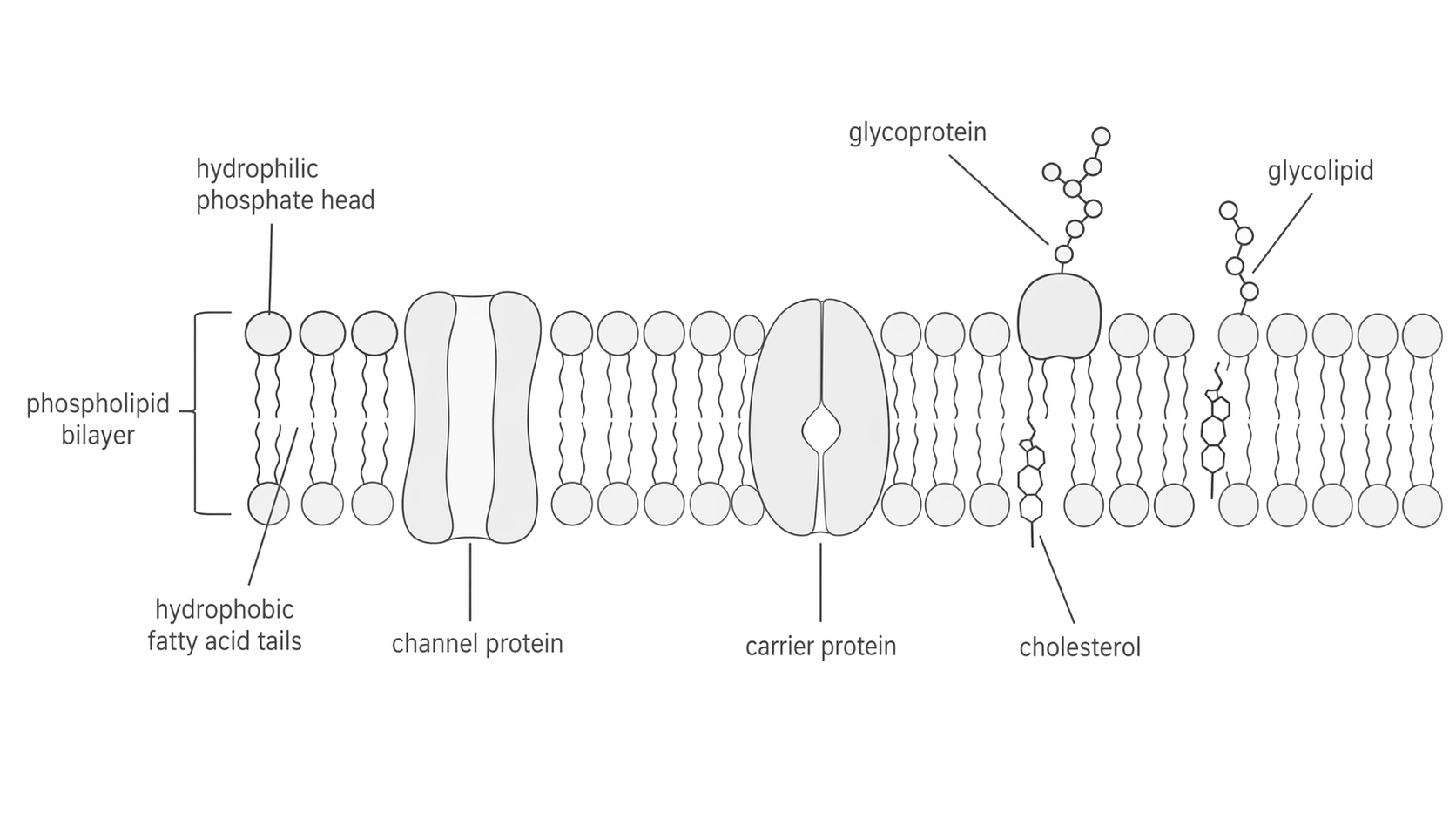
Use the diagram to separate the roles. Channel proteins provide a hydrophilic pore, carrier proteins change shape to move specific substances, glycoproteins and glycolipids project carbohydrate chains for recognition, and cholesterol makes the membrane more stable and less permeable.
Part 2: Diffusion and Facilitated Diffusion
Diffusion is the net movement of particles from a region of higher concentration to a region of lower concentration. Across membranes, the simplest case is small non-polar molecules moving directly through the phospholipid bilayer.
Simple diffusion through the bilayer is suitable for molecules such as oxygen and carbon dioxide. Glucose, amino acids and ions cannot cross this way because they are either too large, too polar or charged.
Facilitated Diffusion
The passive movement of a substance down its concentration gradient through a membrane protein.
Facilitated diffusion uses membrane proteins in two main ways.
- Channel proteins provide a hydrophilic route through the membrane. This is especially important for ions.
- Carrier proteins bind a specific molecule, change shape, and release it on the other side of the membrane.
Neither simple diffusion nor facilitated diffusion requires ATP. In both cases, the substance moves down its concentration gradient.
The rate of diffusion increases when:
- the concentration gradient is steeper
- the membrane surface area is larger
- the diffusion distance is shorter
- the number of suitable channel or carrier proteins is greater
Small non-polar molecules can move through the bilayer itself. Large, polar or charged substances need a protein route.
Part 3: Osmosis and Water Potential
Osmosis is a special case of diffusion because it involves only water.
Osmosis
The net movement of water molecules from a region of higher water potential to a region of lower water potential through a partially permeable membrane.
Water potential is measured in kPa. Pure water has a water potential of 0 kPa. Adding solute lowers water potential, making it more negative. Water therefore moves from a less negative value to a more negative value.
| External water potential compared with the cell | Net water movement | Animal cell | Plant cell |
|---|---|---|---|
| Higher than inside the cell | Into the cell | Swells and may burst | Becomes turgid |
| Equal to inside the cell | No net movement | Remains unchanged overall | No net gain or loss of water overall; the cell is not gaining turgor |
| Lower than inside the cell | Out of the cell | Shrinks / becomes crenated | Loses turgor and may become plasmolysed |
It is worth being precise with plant-cell language. Incipient plasmolysis is the point at which the cell membrane is just beginning to pull away from the cell wall after water loss. It is not simply another way of saying equal water potential.
This is why saline for intravenous drips must be isotonic with blood plasma. If the solution had too high a water potential, red blood cells would take in water and burst.
Part 4: Active Transport and Co-Transport
Some substances must be moved against a concentration gradient. That cannot happen by passive diffusion alone.
Active Transport
The movement of substances across a membrane from a lower concentration to a higher concentration using carrier proteins and energy from ATP hydrolysis.
In active transport:
- The substance binds to a specific carrier protein.
- ATP is hydrolysed to ADP and phosphate.
- The energy released causes the carrier protein to change shape.
- The substance is released on the other side of the membrane.
A classic example is the sodium-potassium pump, which moves sodium ions out of cells and potassium ions into cells.
Co-transport builds on this idea. In the ileum, sodium ions are actively pumped out of epithelial cells into the blood. This keeps the sodium concentration inside the epithelial cell low. Sodium ions then move back in from the gut lumen down their concentration gradient through a co-transporter. Glucose is carried in with them, even when glucose is moving against its own concentration gradient. The glucose then leaves the epithelial cell into the blood by facilitated diffusion.
Co-Transport
Transport in which movement of one substance down its concentration gradient is linked to movement of another substance through the same carrier protein.
This is why oral rehydration solutions contain both glucose and sodium ions: together they help the intestine absorb water more effectively.
The next diagram shows those steps in one ileum epithelial cell. Read it from the lumen side to the blood side and pay attention to where each transport protein is placed: sodium enters with glucose through the co-transporter on the lumen side, sodium is kept low inside the cell by the sodium-potassium pump on the blood side, and glucose then leaves to the blood by facilitated diffusion through a separate carrier.
[DIAGRAM: asset_name: 2.3 - Transport across cell membranes - Diagram 2; asset_slug: 2.3 - Transport across cell membranes - Diagram 2; recommended_method: retained_png; description: Ileum epithelial cell showing sodium-potassium pumps on the blood side, sodium-glucose co-transporters on the lumen side, and glucose leaving to the blood by facilitated diffusion.]
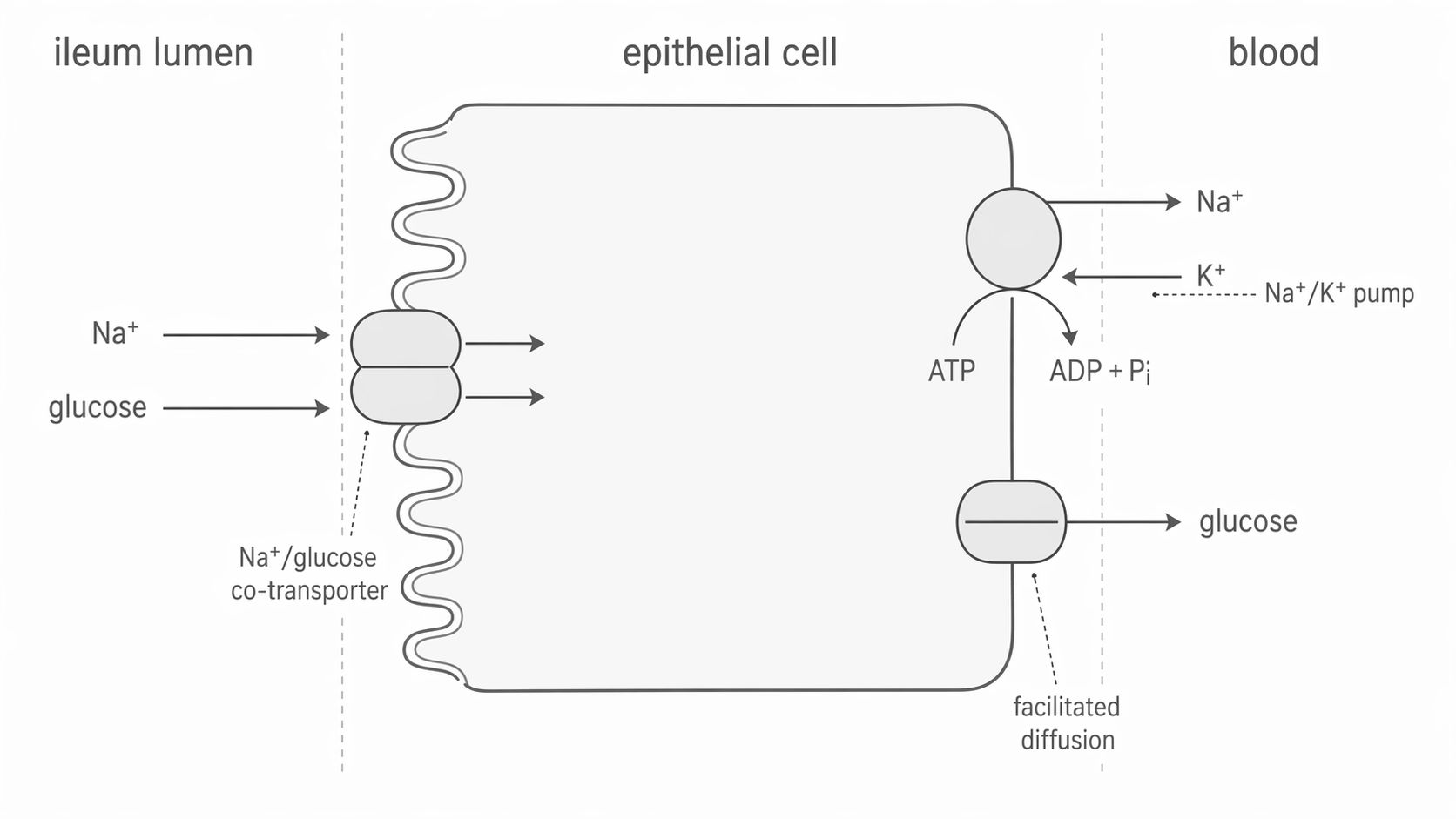
This layout matters because the glucose uptake depends on the sodium gradient. The pump uses ATP to maintain the gradient, the co-transporter uses that gradient to bring glucose into the cell, and the final carrier lets glucose leave into the blood without ATP. If you can explain why the pump must be on the blood side and the co-transporter on the lumen side, you have understood the mechanism.
Part 5: Adaptations for Rapid Transport
Cells that transport substances quickly tend to share the same kinds of adaptations.
| Adaptation | How it increases transport rate | Example |
|---|---|---|
| Large membrane surface area | Gives more area for substances to cross | Microvilli on epithelial cells in the ileum |
| Many channel or carrier proteins | Provides more routes across the membrane | Root hair cells and ileum epithelial cells |
| Steep concentration gradient | Maintains a strong driving force for movement | Blood flow removing absorbed glucose from the ileum |
| Highly folded internal membranes | Gives more space for transport proteins | Cristae in mitochondria and thylakoid membranes in chloroplasts |
If two cells have the same membrane area but one has many more carrier proteins, the cell with more carriers can move suitable substances faster because there are more opportunities for binding and transport.
To explain transport rate, think in three steps: surface area, number of transport proteins, and steepness of the gradient.