3.2.2 - All Cells Arise from Other Cells
This lesson follows genetic material through the cell cycle, mitosis and cytokinesis, linking chromosome behaviour to growth, repair and cancer treatment. It also contrasts eukaryotic cell division with binary fission and with the host-dependent production of virus particles.
Part 1: The Cell Cycle
Every new cell comes from a pre-existing cell. In multicellular organisms, this matters for growth, tissue repair, and the replacement of worn-out cells. Cells that still retain the ability to divide move through a repeating sequence called the cell cycle.
Cell cycle
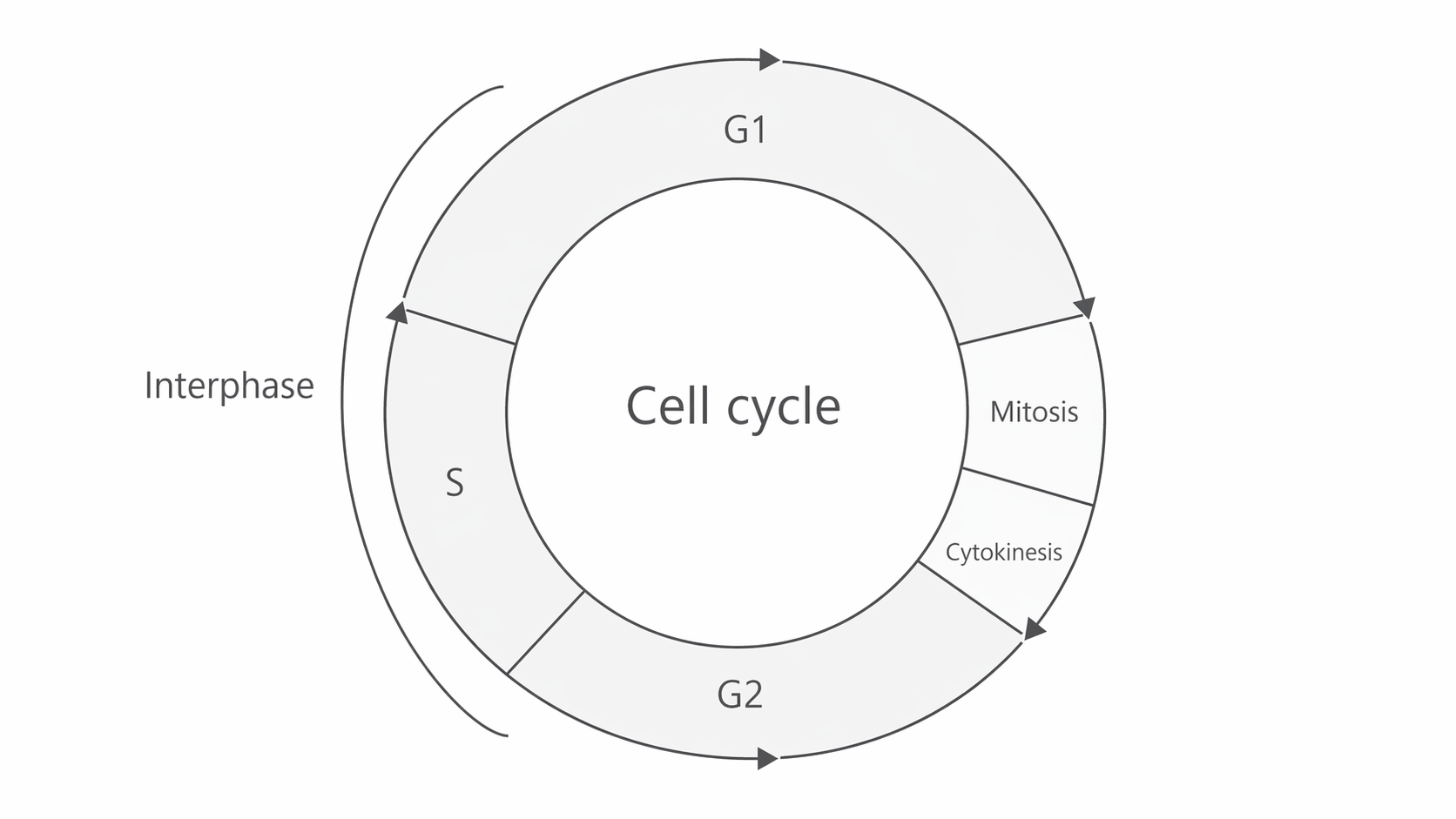
The cell cycle is the sequence of events between one cell division and the next. It includes interphase, mitosis, and usually cytokinesis.
The cell cycle has three main parts:
- Interphase - the cell grows, carries out normal metabolic activity, and prepares for division.
- Mitosis - the nucleus divides.
- Cytokinesis - the cytoplasm divides to form two separate cells.
The diagram below shows the cell cycle as a loop. Notice that interphase takes up most of the circle, and that G1, S, and G2 all sit within this long preparation stage before the much shorter mitosis and cytokinesis stages.
[DIAGRAM: asset_name: 2.2 - All cells arise from other cells - Diagram 1; asset_slug: 2.2 - All cells arise from other cells - Diagram 1; recommended_method: retained_png; description: Circular cell-cycle diagram showing interphase as the largest section, followed by mitosis and cytokinesis. Interphase should be subdivided into G1, S, and G2.]
Interphase
Interphase is not a resting phase. It is a period of intense activity in which the cell prepares for division.
- In G1, the cell grows and synthesises proteins and organelles.
- In S phase, DNA is replicated so each chromosome is copied.
- In G2, the cell continues to grow and synthesises the proteins needed for division.
By the end of interphase, each chromosome consists of two identical sister chromatids joined at the centromere.
In microscope images, chromosomes are not seen as separate dark bodies during interphase because the DNA is uncoiled as chromatin inside an intact nucleus.
Interphase
Interphase is the stage of the cell cycle in which DNA is replicated and the cell prepares for division.
Those preparations involve several distinct cellular processes, which the next check asks you to separate.
Part 2: Mitosis and Cytokinesis
Mitosis
Mitosis is nuclear division that produces two daughter nuclei containing genetic information identical to that of the parent nucleus.
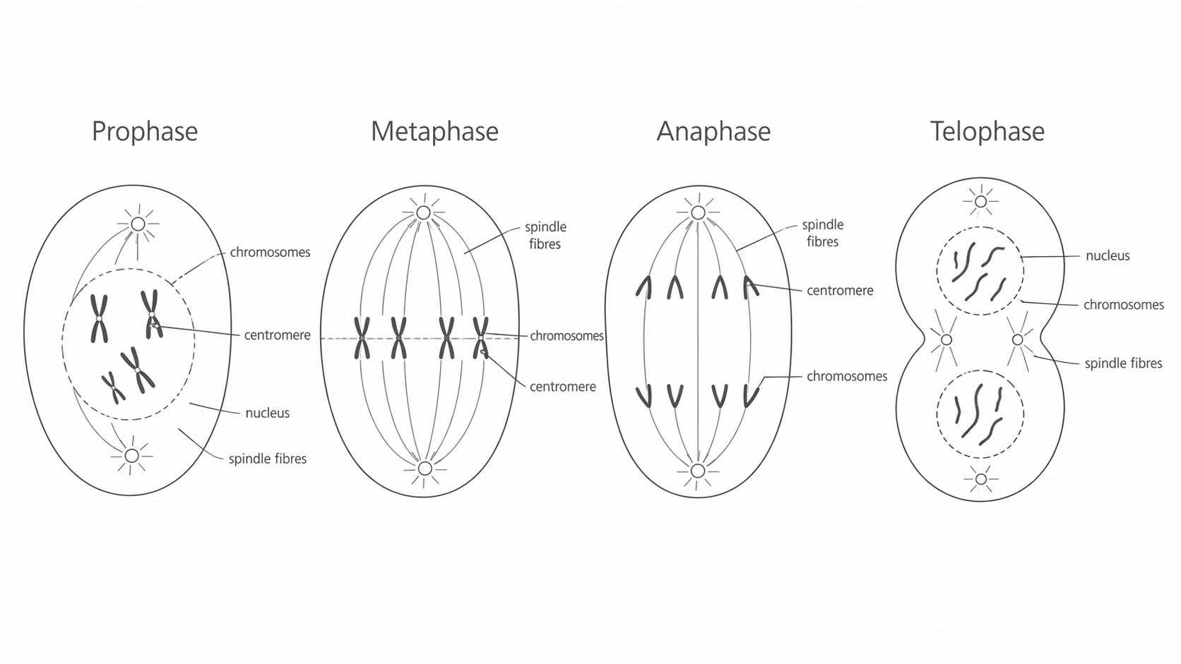
Mitosis is a continuous process, but it is usually described in four stages: prophase, metaphase, anaphase, and telophase.
Prophase
- Chromosomes condense and become visible under the light microscope.
- Each chromosome is made of two sister chromatids joined at the centromere.
- The nuclear envelope breaks down.
- In animal cells, centrioles move to opposite poles and spindle fibres begin to form. Plant cells form spindle fibres without centrioles.
Metaphase
- Chromosomes line up at the equator of the cell.
- Spindle fibres attach to the centromeres.
- This ensures one copy of each chromosome can be moved to each pole.
Anaphase
- The centromeres divide.
- Spindle fibres shorten and pull sister chromatids to opposite poles.
- Once separated, each chromatid is described as a chromosome.
- This movement requires energy from ATP.
Telophase
- Chromosomes reach the poles.
- They uncoil and become less visible.
- A nuclear envelope forms around each group of chromosomes.
- The result is two genetically identical nuclei.
Chromatid
A chromatid is one of two identical copies of a chromosome formed by DNA replication. The two chromatids are joined at the centromere until anaphase.
Recognising Mitotic Stages in a Root Tip Squash
In AQA questions, you are often shown stained cells from a plant root tip and asked to identify the stage from what is visible. Focus on the arrangement of chromosomes and whether the nucleus is intact.
| Stage | What you would see | Best recognition clue |
|---|---|---|
| Interphase | A distinct nucleus is present, but no separate chromosomes can be seen. The DNA is spread out as chromatin. | The nucleus looks intact and the chromosomes are not individually visible. |
| Prophase | Chromosomes first become visible as dark, condensed threads or clumps. The nuclear envelope is breaking down. | You can see condensed chromosomes, but they are not lined up across the middle yet. |
| Metaphase | Chromosomes are arranged across the equator of the cell. | The chromosomes form a clear line across the middle of the cell. |
| Anaphase | Sister chromatids have separated and are moving to opposite poles. | Two groups of chromosomes are being pulled apart, often appearing V-shaped. |
| Telophase | Chromosomes have reached the poles and start to uncoil. Two nuclei begin to form. | There are two chromosome groups at opposite ends and two new nuclei may be visible. |
| Cytokinesis | The cytoplasm is dividing. In plant cells, a cell plate forms across the middle. | In root tip cells, look for a developing cell plate between the two new nuclei. |
One helpful exam clue is that interphase is usually the most common stage in a root tip squash because cells spend much longer in interphase than in mitosis.
The next diagram summarises the four mitotic stages in order. As you study it, track how the chromosomes condense, line up, separate, and then become enclosed in new nuclei, and notice exactly where the spindle fibres attach and what happens at the centromere in each panel.
[DIAGRAM: asset_name: 2.2 - All cells arise from other cells - Diagram 2; asset_slug: 2.2 - All cells arise from other cells - Diagram 2; recommended_method: retained_png; description: Four-panel sequence of mitosis showing prophase, metaphase, anaphase, and telophase in a eukaryotic cell. Label chromosomes, centromeres, spindle fibres, and nuclei.]
Cytokinesis
After mitosis, the cytoplasm divides.
- In animal cells, the cell-surface membrane pinches inward.
- In plant cells, vesicles form a cell plate that develops into a new cell wall.
This produces two separate daughter cells.
Part 3: Why Mitosis Matters
Mitosis is important because it produces genetically identical cells.
- Growth - a zygote divides repeatedly to produce the many cells of a multicellular organism.
- Repair and replacement - damaged or worn-out cells can be replaced with cells that have the same genes.
- Asexual reproduction - some organisms produce offspring by mitosis, so the offspring are genetically identical to the parent apart from any mutation.
Genetic similarity matters because replacement cells must usually make the same proteins and carry out the same function as the cells they replace.
Part 4: Control of Cell Division and Cancer
Cell division is controlled by genes and cell-signalling molecules. If control is lost, cells may divide too often and form a tumour.
Tumour
A tumour is a mass of abnormal cells produced by uncontrolled cell division.
Tumours may be:
- Benign - contained in one place and not invasive.
- Malignant - invasive and able to spread to other parts of the body.
Many cancer treatments target cells that are dividing rapidly.
- Some drugs stop DNA replication.
- Some drugs stop spindle fibres forming, so chromosomes cannot separate properly.
These treatments can also affect normal tissues in which cells divide frequently, such as the gut lining, bone marrow, and hair follicles.
Part 5: Binary Fission in Prokaryotes
Binary fission
Binary fission is the way prokaryotic cells divide to produce two daughter cells.
Prokaryotes do not divide by mitosis because they do not have a nucleus or linear chromosomes. Instead, they divide by binary fission.
- The circular DNA replicates.
- Plasmids replicate independently.
- The cell divides so that each daughter cell receives one copy of the circular DNA and a variable number of plasmids.
Unlike mitosis, binary fission does not involve chromosome condensation, spindle fibres, or a nuclear envelope.
Part 6: Why Viruses Do Not Divide
Virus
A virus is an acellular particle made of nucleic acid surrounded by a protein coat. It can replicate only inside a host cell.
Viruses are non-living and do not undergo cell division.
Instead:
- A virus attaches to a host cell.
- Its nucleic acid enters the host cell.
- The host cell then uses its own enzymes, ribosomes, ATP, and raw materials to make viral nucleic acid and viral proteins.
- New virus particles are assembled.
- The new viruses leave the host cell, often when the cell lyses.
This is why the specification states that, after injection of viral nucleic acid, the infected host cell replicates the virus particles.
Quick Recap
The lesson describes three different routes to new cells or particles. Compare what happens to genetic material in each route.