3.1.4.1 - General Properties of Proteins
This lesson traces proteins from the common structure of amino acids to peptide bonds, polypeptides and the four levels of protein structure. It connects interactions between R groups to three-dimensional shape and function, then uses the Biuret test to detect peptide bonds.
Part 1: Amino Acids
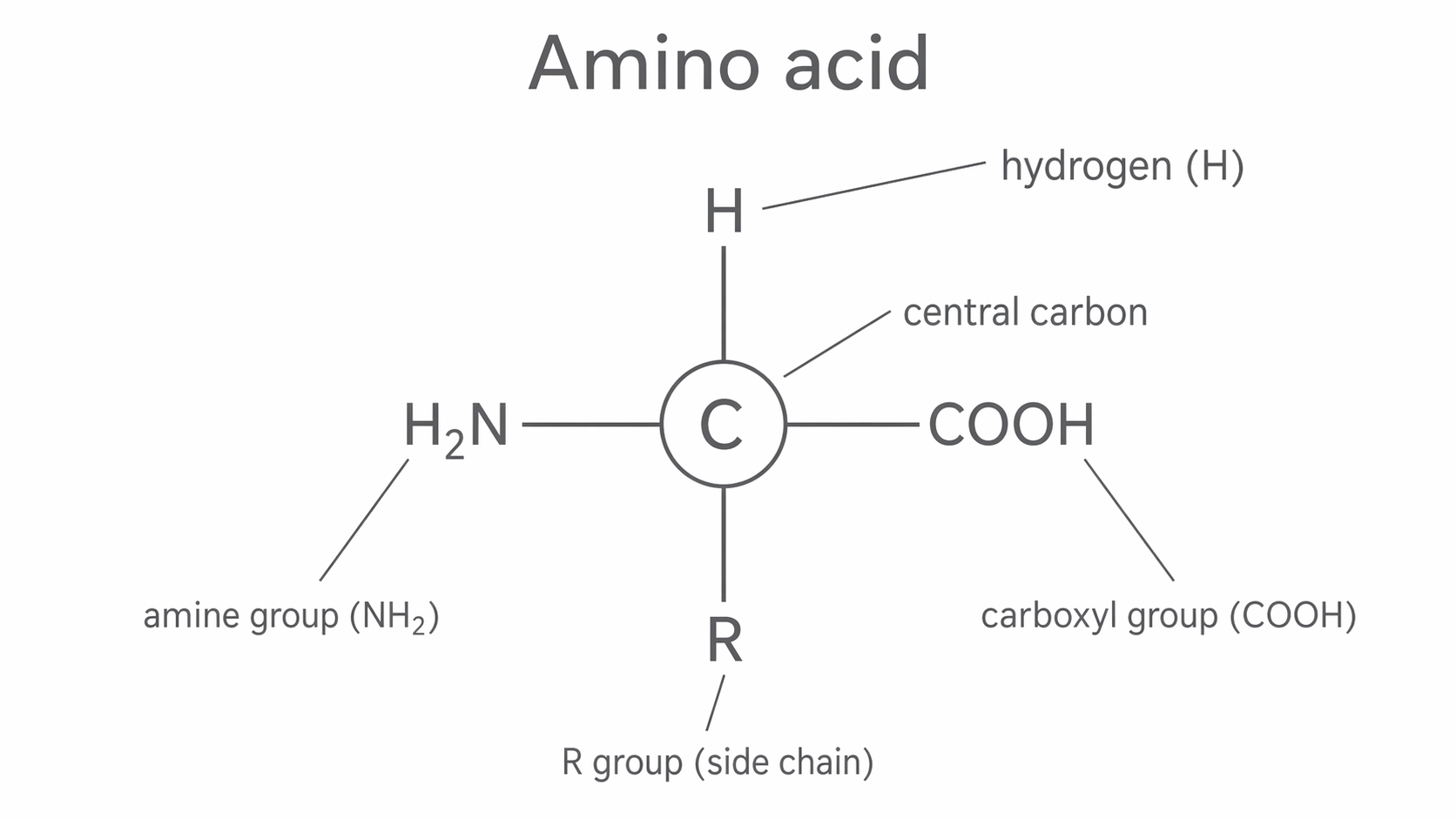
Proteins are made from amino acid monomers. Every amino acid has the same basic layout: a central carbon atom bonded to an amine group (NH2), a carboxyl group (COOH), a hydrogen atom, and an R group. The R group is the side chain, and it is the only part that differs between the twenty amino acids common to living organisms.
That shared structure matters because it lets amino acids join together in the same general way, while the different R groups give different amino acids different chemical properties. Some side chains are charged, some are polar, and some contain sulfur. Those differences help determine how a protein folds and therefore what it can do.
In the diagram below, notice that the central carbon and the NH2, COOH, and H groups stay the same in every amino acid. The only variable part is the R group, which is why biologists focus on side chains when comparing amino acids.
[DIAGRAM: asset_name: 1.4.1 - General properties of proteins - Diagram 1; asset_slug: 1.4.1 - General properties of proteins - Diagram 1; recommended_method: retained_png; description: General structure of an amino acid showing the central carbon bonded to NH2, COOH, H, and R, with each group labelled.]
Part 2: Peptide Bonds and Polypeptides
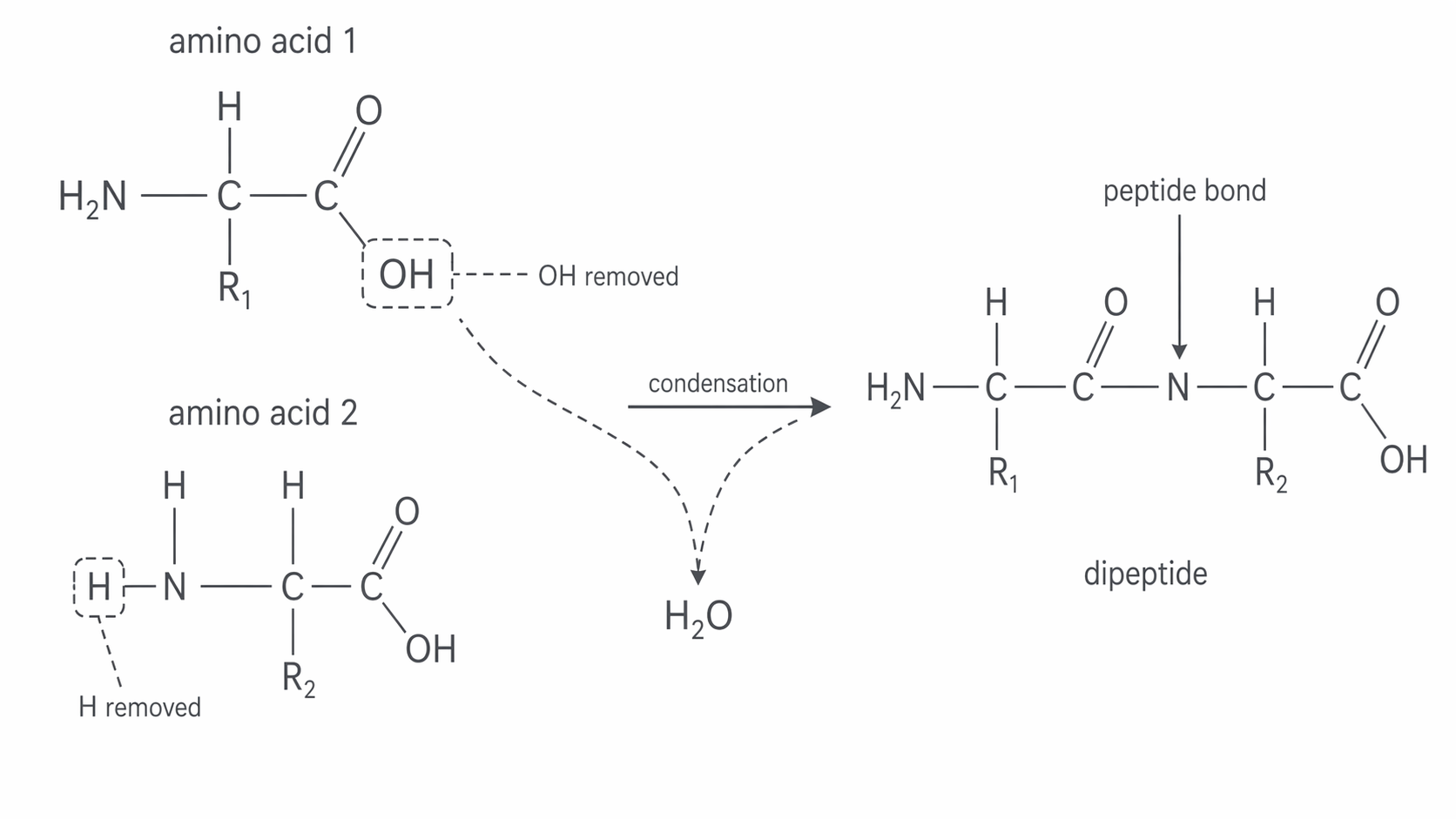
When two amino acids join, the carboxyl group of one reacts with the amine group of the other. A molecule of water is removed, so this is a condensation reaction. The new covalent bond formed between the two amino acids is called a peptide bond.
Two amino acids joined together form a dipeptide. A long chain of many amino acids joined by peptide bonds is a polypeptide. Some proteins are made from a single polypeptide chain, while others contain two or more polypeptide chains linked together.
The reverse process is hydrolysis. In hydrolysis, water is added to break peptide bonds. This matters biologically because proteins in food are digested by hydrolysis before their amino acids can be absorbed.
In the diagram below, trace the OH removed from one amino acid and the H removed from the other, then identify the peptide bond left behind.
[DIAGRAM: asset_name: 1.4.1 - General properties of proteins - Diagram 2; asset_slug: 1.4.1 - General properties of proteins - Diagram 2; recommended_method: retained_png; description: Two amino acids joining by condensation, showing removal of OH from one amino acid and H from the other to make water, and the peptide bond left behind.]
Part 3: Levels of Protein Structure
The properties of a protein depend on its structure at several levels.
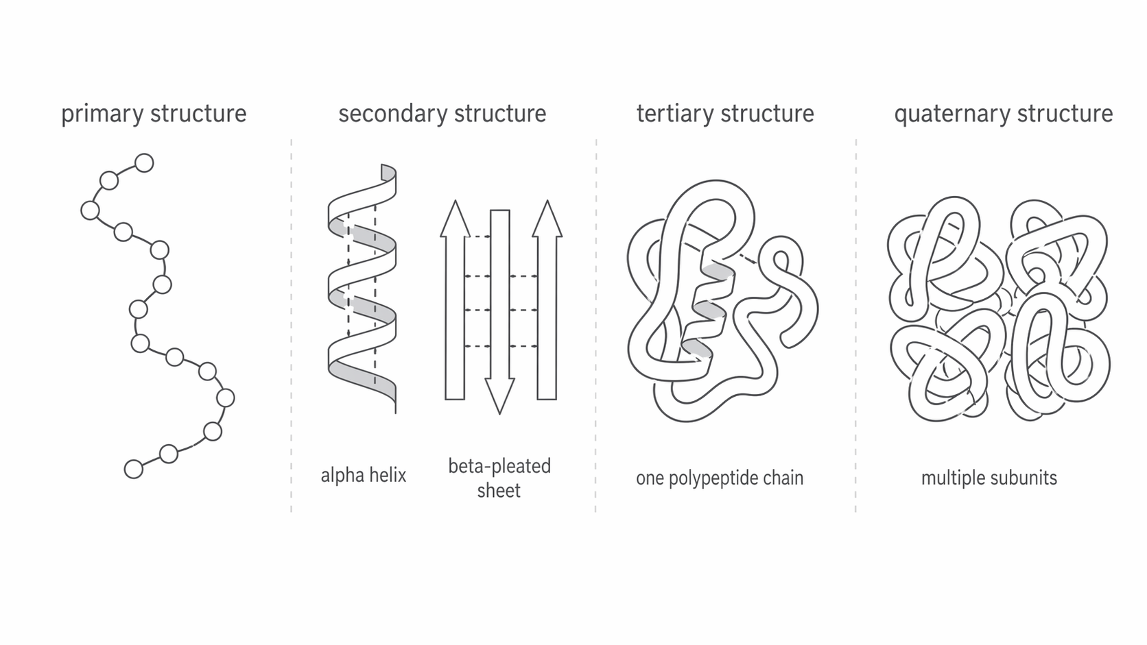
The primary structure is the sequence of amino acids in the polypeptide chain. This sequence is crucial because it determines all higher levels of structure. Even one amino acid change can alter the final shape of the protein and reduce or change its function.
The secondary structure is the regular folding of the polypeptide chain into shapes such as an alpha helix or a beta-pleated sheet. These shapes are held in place by hydrogen bonds between the NH and C=O groups in the backbone of the polypeptide.
The tertiary structure is the overall three-dimensional shape of one polypeptide chain. This shape is maintained by interactions between side chains, including:
- hydrogen bonds
- ionic bonds
- disulfide bridges
Disulfide bridges form between sulfur-containing side chains, especially between cysteine residues, and help stabilise the folded protein.
The quaternary structure is the way two or more polypeptide chains are arranged to make the final functional protein. Haemoglobin is a good example because it contains four polypeptide subunits and works only when those subunits are assembled correctly.
In the diagram below, compare the scale of each level carefully: primary is just the amino acid sequence, secondary shows local backbone folding, tertiary is the full 3D shape of one polypeptide, and quaternary is the arrangement of multiple polypeptide subunits.
[DIAGRAM: asset_name: 1.4.1 - General properties of proteins - Diagram 3; asset_slug: 1.4.1 - General properties of proteins - Diagram 3; recommended_method: retained_png; description: Protein structure overview showing primary sequence, secondary alpha helix/beta-pleated sheet, tertiary folded polypeptide, and quaternary arrangement of multiple subunits.]
Part 4: Structure and Function
Protein function depends on shape. Globular proteins fold into compact shapes and are usually soluble, which suits roles such as catalysis and transport. Enzymes and haemoglobin are globular proteins.
Fibrous proteins are long, strong, and usually insoluble, which suits structural roles. Collagen is an important example. It contains three polypeptide chains wound around each other, forming a strong rope-like molecule. Cross-links between the chains help make collagen tough and resistant to stretching, which is why it is well suited to tendons and other connective tissues.
The key idea for this topic is simple: the amino acid sequence determines how a protein folds, and the final shape determines how the protein behaves. If the structure changes, the function usually changes as well.
Part 5: The Biuret Test
The Biuret test is used to test for proteins because it detects peptide bonds.
The method is:
- Add sodium hydroxide solution to the sample to make the mixture alkaline.
- Add a few drops of dilute copper(II) sulfate solution, or simply add Biuret reagent.
- A purple colour shows that peptide bonds are present.
- If no protein is present, the solution stays blue.
This is a qualitative test. It tells you whether protein is present, not how much protein there is.
Keep the outcomes paired clearly in your memory: a negative result stays blue, while a positive result turns purple because peptide bonds are present.