3.1.4.2 - Many Proteins Are Enzymes
Enzymes connect the precise tertiary structure of proteins to the control of biological reactions. This lesson develops models of enzyme action, explains how conditions and concentrations affect rate, and compares the ways inhibitors regulate enzyme-controlled pathways.
Part 1: Enzymes as Catalysts
Many proteins are enzymes. Enzymes are biological catalysts, which means they increase the rate of a chemical reaction without being used up in the process. They make reactions happen fast enough for life by lowering the activation energy needed for the reaction to start.
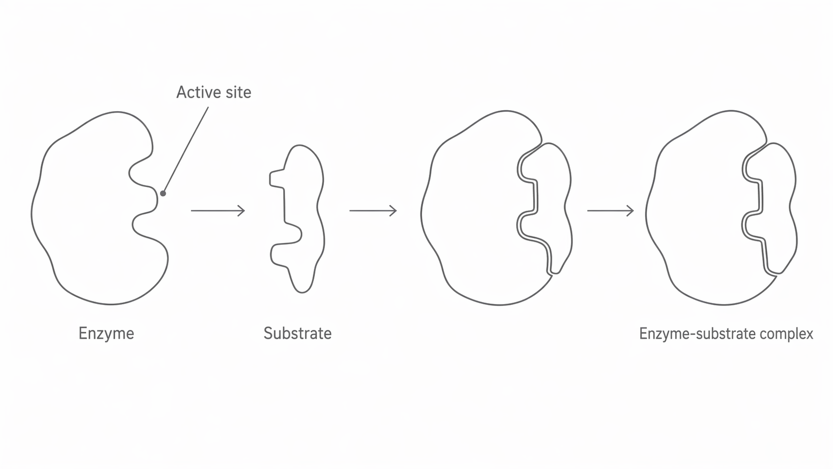
Enzymes are globular proteins with a precise tertiary structure. Part of that structure forms the active site, a small region where the substrate binds. The shape of the active site is complementary to the substrate, not identical to it. When a substrate binds, an enzyme-substrate complex forms.
Because the active site depends on tertiary structure, small changes in shape can change how well the substrate binds. This is why enzymes are usually highly specific: each enzyme normally catalyses one reaction, or a narrow range of closely related reactions.
In the diagram below, notice that the substrate is complementary to the active site rather than identical to it. Then follow the sequence from separate enzyme and substrate to the enzyme-substrate complex formed when binding occurs.
[DIAGRAM: asset_name: 1.4.2 - Many proteins are enzymes - Diagram 1; asset_slug: 1.4.2 - Many proteins are enzymes - Diagram 1; recommended_method: retained_png; description: Enzyme with a labelled active site binding a complementary substrate to form an enzyme-substrate complex.]
Part 2: Models of Enzyme Action
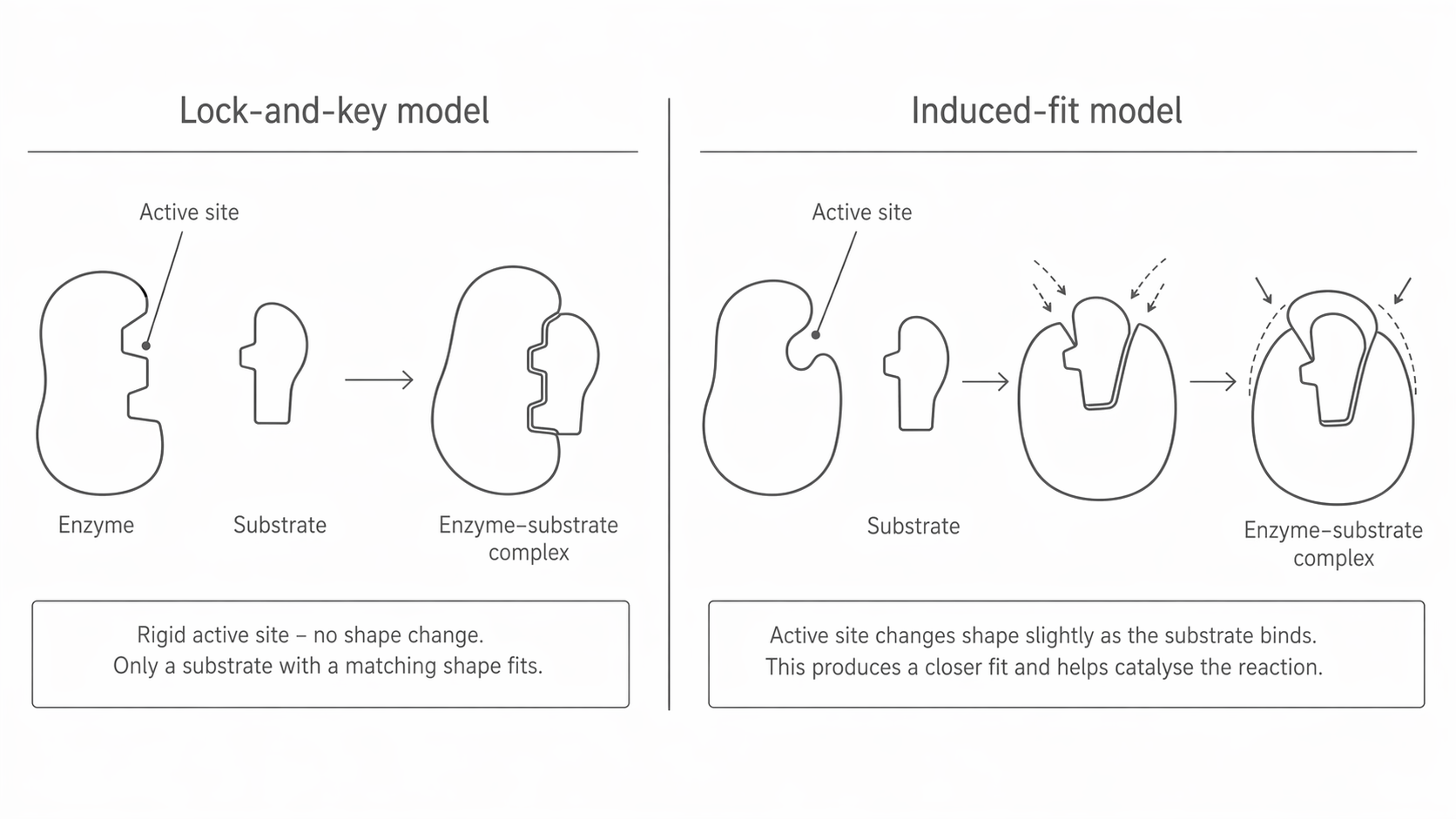
Early scientists used the lock and key model to explain enzyme specificity. In that model, the active site is treated as a rigid shape that fits only one substrate, like a lock fits one key. This helps explain why enzymes are specific, but it does not explain all observations.
The model now used in A-level biology is the induced fit model. In this model, the active site is flexible. The substrate approaches an active site that is already broadly complementary, and binding causes the enzyme to change shape slightly so that the fit becomes closer. This conformational change can place strain on bonds in the substrate, helping the reaction happen and lowering activation energy.
The shift from lock and key to induced fit is a good example of how scientific models change. A model is useful only while it explains the evidence. When new observations show that the old model is incomplete, scientists refine or replace it.
Use the diagram below to compare the two models directly. The key detail to notice is whether the active site stays rigid in the lock and key model or changes shape around the substrate in the induced fit model.
[DIAGRAM: asset_name: 1.4.2 - Many proteins are enzymes - Diagram 2; asset_slug: 1.4.2 - Many proteins are enzymes - Diagram 2; recommended_method: retained_png; description: Two-panel comparison showing a rigid lock-and-key model on one side and an induced-fit model with the active site changing shape on the other.]
Part 3: Temperature and pH
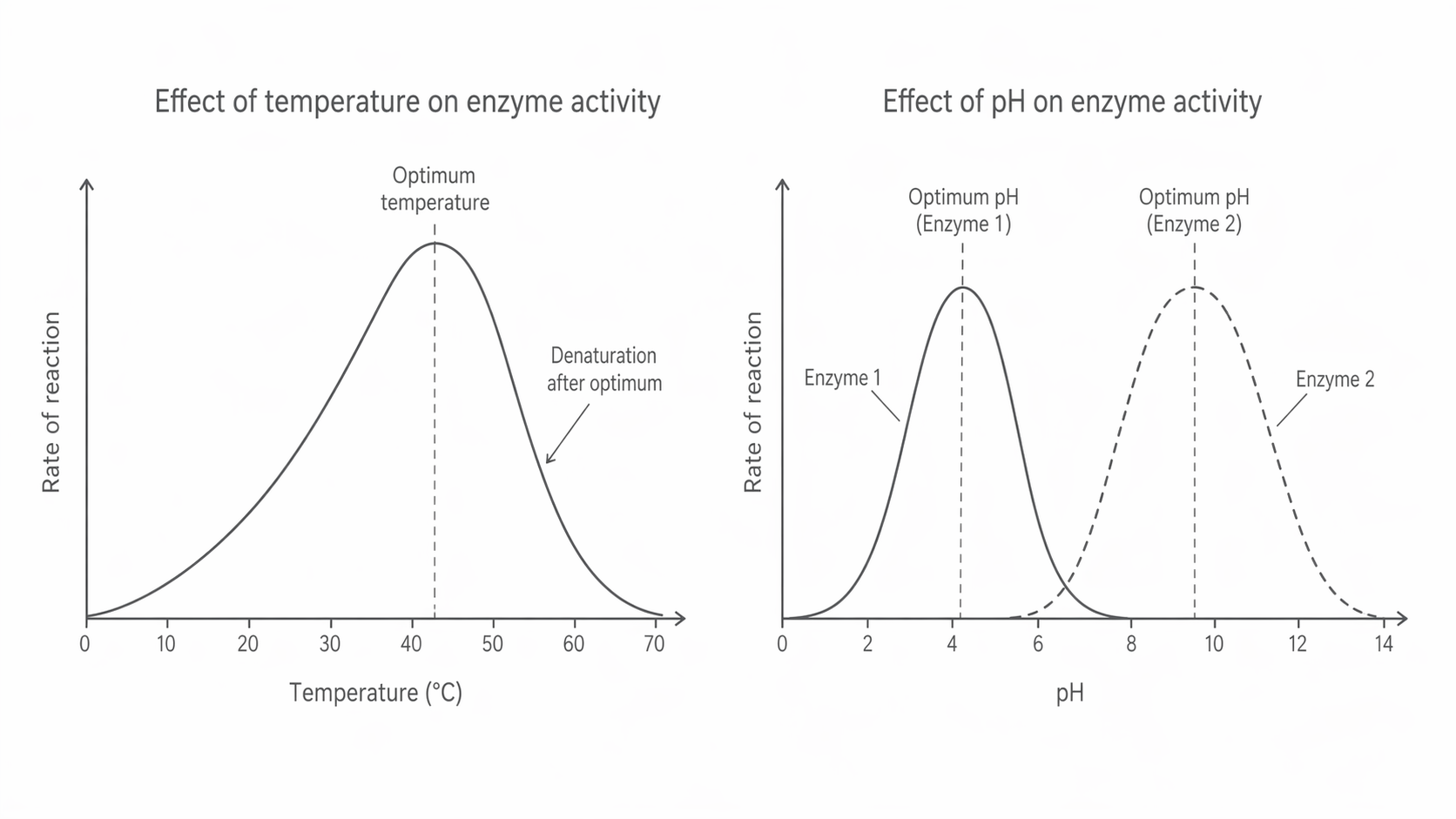
At low temperatures, enzyme and substrate molecules have less kinetic energy. They move more slowly, so there are fewer successful collisions per second and the rate of reaction is low. Low temperature does not usually denature the enzyme; it simply slows the reaction.
As temperature rises, molecules move faster and collide more often. More enzyme-substrate complexes form each second, so the rate increases up to an optimum temperature.
Above the optimum, heat disrupts bonds that help maintain the enzyme's tertiary structure, especially hydrogen bonds and ionic bonds. The active site changes shape, the substrate no longer fits as well, and the rate falls. If the change is large enough, the enzyme becomes denatured and can no longer catalyse the reaction.
pH also affects enzyme shape and function. Changes in H+ concentration can alter the charges on amino acid side chains, especially around the active site. That can disrupt ionic bonds and hydrogen bonds, change the active site's shape, and reduce the rate. Each enzyme has an optimum pH, and moving away from it lowers activity. Extreme pH can denature the enzyme.
Different enzymes therefore work best under different conditions. For example, pepsin works best in the acidic conditions of the stomach, while enzymes in the small intestine work best in more alkaline conditions.
The graphs below are there to help you read the shape of the relationship, not just memorise the words. Notice the optimum peak for temperature, the sharp drop when denaturation changes the active site, and the fact that different enzymes can have different pH optima.
[DIAGRAM: asset_name: 1.4.2 - Many proteins are enzymes - Diagram 3; asset_slug: 1.4.2 - Many proteins are enzymes - Diagram 3; recommended_method: retained_png; description: One graph of rate against temperature with an optimum peak, and one graph of rate against pH for two different enzymes with different optima.]
Part 4: Enzyme Concentration and Substrate Concentration
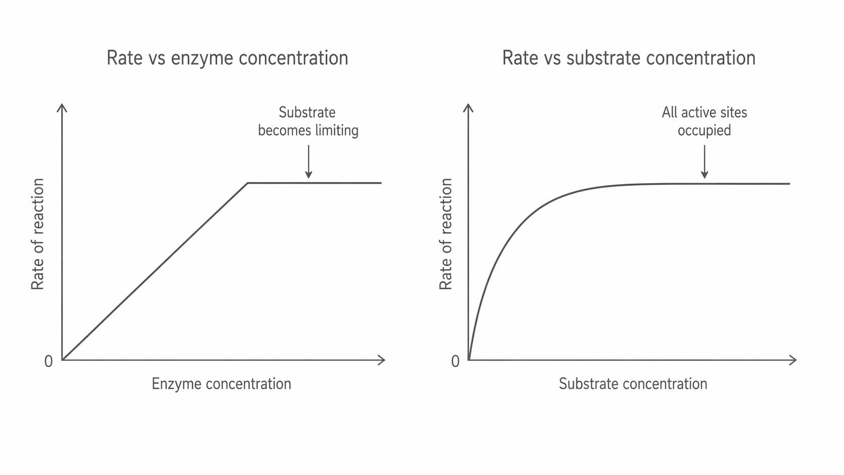
If substrate concentration is kept high, increasing enzyme concentration increases the rate of reaction because more active sites are available. More enzyme-substrate complexes can form each second. This increase continues only while substrate is not limiting. Once substrate becomes the limiting factor, adding more enzyme does not increase the rate further.
If enzyme concentration is kept constant, increasing substrate concentration increases the rate at first because substrates collide with active sites more often. Eventually, all active sites become occupied as fast as they are available. At that point the enzyme is working at its maximum rate, so adding more substrate has no further effect.
These two factors show the same general principle: the rate rises until some other factor becomes limiting.
The graphs below show that shared pattern clearly: the rate rises at first, then levels off once some other factor becomes limiting. Notice that both curves plateau, but for different reasons: substrate becomes limiting in one graph, while all active sites are occupied in the other.
[DIAGRAM: asset_name: 1.4.2 - Many proteins are enzymes - Diagram 4; asset_slug: 1.4.2 - Many proteins are enzymes - Diagram 4; recommended_method: retained_png; description: One graph of rate against enzyme concentration, rising then levelling off when substrate becomes limiting, and one graph of rate against substrate concentration, rising then levelling off when all active sites are occupied.]
Part 5: Enzyme Inhibition
An enzyme inhibitor is a substance that reduces the rate of an enzyme-controlled reaction.
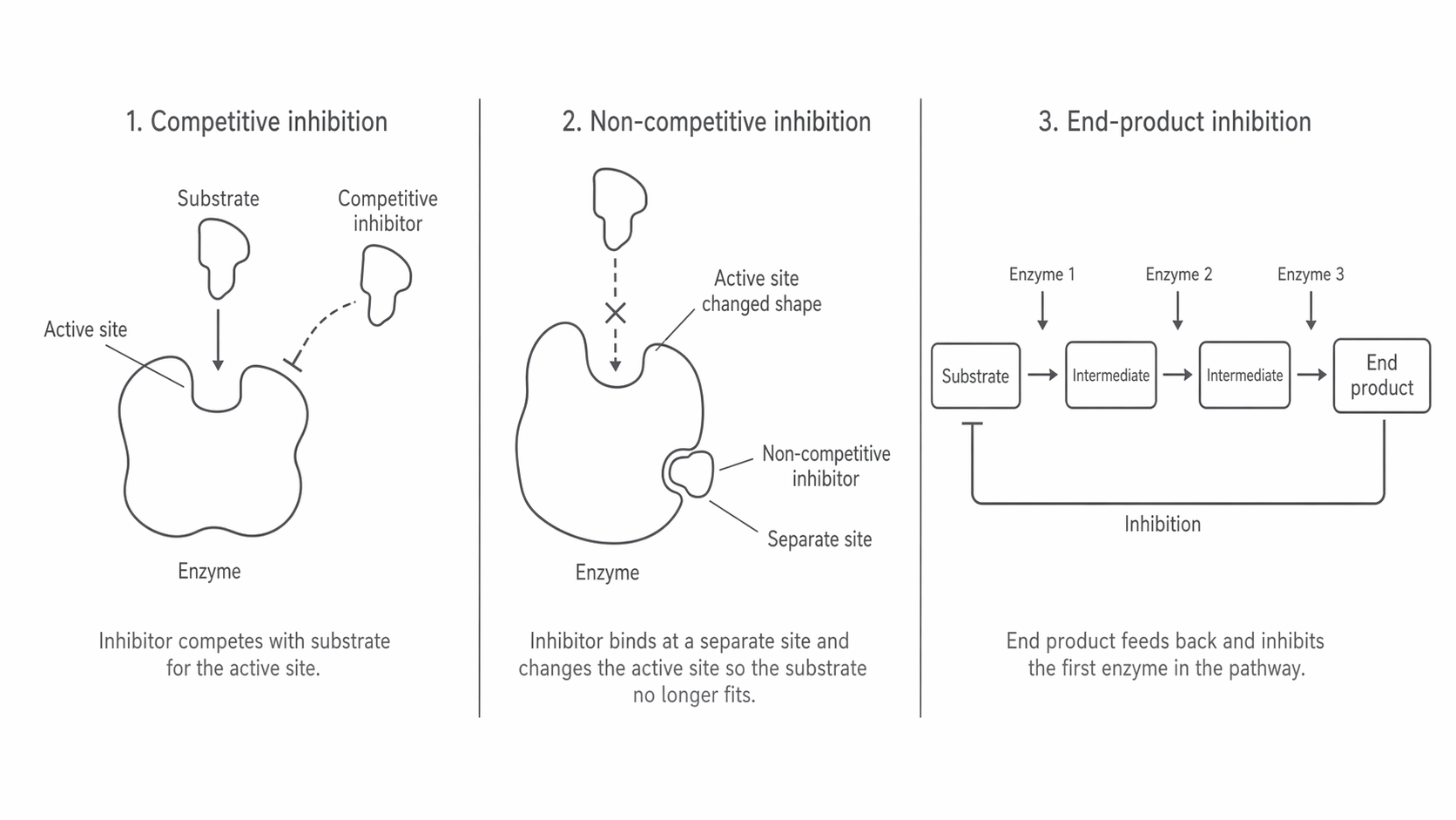
A competitive inhibitor has a shape similar to the substrate and competes for the active site. As the concentration of competitive inhibitor increases, more active sites are blocked at any one moment, so the rate falls. Its effect can be reduced by increasing substrate concentration because substrate molecules then have a better chance of reaching the active site first. A safe textbook example is malonate, which competes with succinate for the active site of succinate dehydrogenase.
A non-competitive inhibitor binds to a different site on the enzyme, not the active site. This changes the enzyme's shape and alters the active site so the substrate no longer fits properly. As the concentration of non-competitive inhibitor increases, more enzyme molecules are affected and the rate falls further. Increasing substrate concentration does not overcome this effect because the substrate is not competing for the same binding site.
Cells use this idea in end-product inhibition. In some metabolic pathways, the final product acts as a non-competitive inhibitor of an enzyme near the start of the pathway. If the end product builds up, it slows further production. If its concentration falls, inhibition is reduced and production increases again.
The diagram below is useful because it lets you compare all three cases at once. Notice where each inhibitor binds, how non-competitive inhibition changes the active site indirectly, and how the final product in a pathway can feed back to reduce further production.
[DIAGRAM: asset_name: 1.4.2 - Many proteins are enzymes - Diagram 5; asset_slug: 1.4.2 - Many proteins are enzymes - Diagram 5; recommended_method: retained_png; description: Competitive inhibition at the active site, non-competitive inhibition at a separate site, and a simple pathway showing end-product inhibition.]
Part 6: Enzymes in Living Systems
Enzymes catalyse reactions both inside cells and outside cells. Intracellular enzymes take part in processes such as respiration, photosynthesis, DNA replication, and protein synthesis. Extracellular enzymes include digestive enzymes such as amylase and trypsin, which are released from cells and work outside them.
Together, enzymes control metabolism from the cellular level to the whole organism. In some cases, enzymes in a pathway are associated with particular organelles or membranes, which can help organise a sequence of reactions. In many other cases, they simply work in the cytoplasm or in the fluid inside organelles. Either way, the product of one enzyme-controlled reaction often becomes the substrate for the next.
This is why enzyme action matters so much in biology: enzymes do not just speed up isolated reactions, they organise whole systems of life.