4.2.2 - Brain localisation, lateralisation, plasticity and recovery
Some brain functions are concentrated in specialised areas, yet experience and injury can reorganise the networks that support them. You will locate motor, somatosensory, visual, auditory and language functions, examine hemispheric lateralisation through split-brain research, and explain neuroplasticity and functional recovery after trauma. Evidence from cases and scanning methods supports evaluation of strict localisation, individual variation and limits on recovery.
Localisation and lateralisation
This lesson is about two linked ideas. First, some psychological and behavioural functions are associated with particular brain areas. Second, the two hemispheres are not identical: some functions are more strongly associated with one hemisphere than the other.
Localisation of function
Localisation of function is the idea that different areas of the brain are responsible for different functions, such as movement, touch, vision, hearing and language.
Hemispheric lateralisation
Hemispheric lateralisation means that a function is mainly controlled by, or more strongly associated with, one hemisphere of the brain.
The strongest answer does not treat the brain as a set of separate switches. Localised areas matter, but most behaviours depend on networks of areas communicating with each other. For example, speaking a sentence may involve language planning, motor control of the mouth, hearing your own voice, memory, attention and emotion.
[DIAGRAM: asset_name: Localised cortical centres; asset_slug: 4_2_2_06_localisation_of_function_in_the_brain_and_hemispheric_lateralisation__diagram_01; recommended_method: image_gen; description: Simplified left lateral cerebral cortex showing frontal, parietal, visual, auditory, motor, somatosensory, Broca's and Wernicke's areas, with a note that areas are approximate rather than isolated switches.]
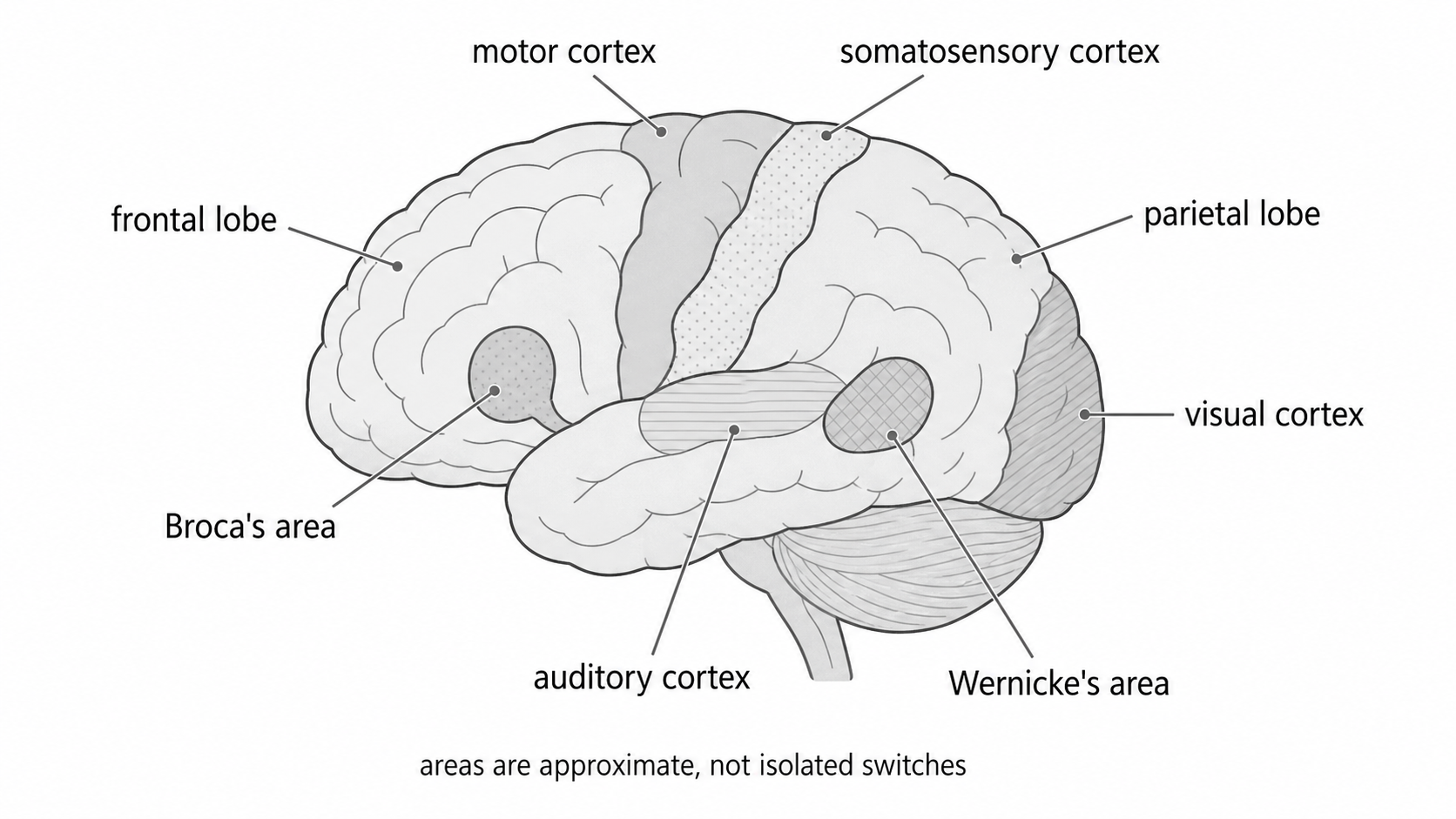
The required functions
The main areas and processes are:
- motor centre: controls voluntary movement;
- somatosensory centre: processes touch, pressure, pain and body-position information;
- visual centre: processes visual information;
- auditory centre: processes sound;
- language centres, especially Broca's area and Wernicke's area;
- split-brain research, used as evidence for hemispheric lateralisation;
- plasticity and functional recovery after brain trauma.
Using the idea in context
If a person has damage to the back of the brain and develops visual difficulties, a localisation explanation would link the difficulty to the visual cortex in the occipital lobe. If a person understands speech but struggles to produce fluent speech after damage to the left frontal lobe, a localisation explanation would link this to Broca's area.
First caution
Localisation is useful because it helps psychologists and clinicians predict symptoms from damage and design investigations. The caution is that complex behaviours are rarely controlled by one tiny area alone. A sophisticated answer says localised contribution, not single-area cause.
Motor, somatosensory, visual and auditory centres
The four cortical centres make different contributions, with movement and body sensation mainly organised contralaterally.
Motor centre
The motor cortex is in the frontal lobe, just in front of the central sulcus. It is involved in voluntary movement. The control is largely contralateral, meaning the left motor cortex controls movement on the right side of the body and the right motor cortex controls movement on the left side.
The motor cortex is organised roughly by body part. Areas requiring fine control, such as the hands and mouth, have a larger representation than areas requiring less precise control. You do not need to draw a motor homunculus for this lesson, but the principle is useful: movement control is localised and mapped.
Somatosensory centre
The somatosensory cortex is in the parietal lobe, just behind the central sulcus. It processes body sensation, including touch, pressure and body-position information, and contributes to processing pain and temperature alongside other pathways and areas. Like the motor cortex, it is mostly contralateral: information from the right side of the body is processed mainly in the left somatosensory cortex, and vice versa.
Visual centre
The visual cortex is in the occipital lobe at the back of the brain. It processes visual information from the eyes. Information from the left visual field is processed mainly by the right hemisphere, and information from the right visual field is processed mainly by the left hemisphere. Each visual field includes information from both eyes; it is not the same as one eye. This distinction matters in split-brain research.
Auditory centre
The auditory cortex is in the temporal lobe. It processes sound information, including features such as pitch, rhythm and loudness. Auditory processing is more bilateral than the simple "left side/right side" pattern used for body movement, so avoid claiming that only one auditory cortex processes one ear.
Symptom matching
Imagine these patients:
- Maya has damage just in front of the central sulcus in the left hemisphere and finds it hard to move her right hand deliberately. This fits damage to the motor cortex.
- Rafi can move his hand but cannot feel pressure or temperature from it normally. This fits damage to the somatosensory cortex.
- Ella has damage at the back of the brain and has problems processing visual information. This fits damage to the visual cortex.
- Noah has temporal-lobe damage and difficulty processing sounds. This fits damage around the auditory cortex.
Useful but simplified
These centres give strong evidence for localisation because damage to different areas tends to produce different symptoms. However, localisation can be oversimplified. Vision, hearing and movement all involve pathways before and after the cortical centre, so a symptom may come from damage to a pathway, not only the named cortex.
Language centres: Broca and Wernicke
Language illustrates both localisation and lateralisation. In most people, especially most right-handed people, language functions are strongly associated with the left hemisphere. This does not mean the right hemisphere does nothing for language; it can contribute to tone, emotion, context and broader interpretation.
Aphasia
Aphasia is an impairment of language caused by brain damage. It may affect speech production, comprehension, reading or writing.
Broca's area
Broca's area is usually described as being in the left frontal lobe, near areas involved in mouth and tongue movement. It is associated with speech production.
Damage to Broca's area is linked to Broca's aphasia, sometimes called expressive aphasia. A person may understand more than they can produce, but their speech is slow, effortful and lacking grammatical fluency. They may know what they want to say but struggle to get the words out smoothly.
Classic evidence came from Paul Broca's 19th-century clinical observations of patients with severe speech production difficulties and damage in the left frontal region. The historical cases were important because they suggested that speech production was not spread evenly across the whole brain.
Wernicke's area
Wernicke's area is usually described as being in the left posterior temporal region, close to auditory-processing areas. It is associated with language comprehension.
Damage to Wernicke's area is linked to Wernicke's aphasia, sometimes called receptive aphasia. A person may speak fluently, but the speech may lack meaning, include incorrect words, or fail to answer the question asked. They may also struggle to understand spoken or written language.
Carl Wernicke's 1874 work supported the idea that language involved more than one centre. Production and comprehension could be impaired differently, suggesting partial localisation within the language system.
Applying aphasia
If Amira says, "I... want... shop... tomorrow," and appears frustrated because she understands the question but cannot speak fluently, Broca's aphasia is more likely. If Denzel speaks smoothly but says sentences that do not make sense and cannot follow simple verbal instructions, Wernicke's aphasia is more likely.
Modern caution
Broca's and Wernicke's areas are useful parts of a model, while modern neuroscience treats language as a network. Dronkers et al. (2007) scanned the preserved brains of Broca's two historic patients. Their lesions extended into deeper regions as well as the surface damage Broca observed. The original speech difficulties therefore cannot be attributed confidently to that surface area alone.
This qualifies strict localisation rather than showing that location is irrelevant. A lesion may disrupt a specialised area, its connections, or both. Likewise, a fluent but meaningless speech pattern in a brief scenario supports a Wernicke-type interpretation, rather than providing enough information for a clinical diagnosis.
Split-brain research and hemispheric lateralisation
Split-brain research studies people whose corpus callosum has been surgically cut, usually as a treatment for severe epilepsy. The corpus callosum is the bundle of fibres that normally allows the two hemispheres to share information.
Split-brain research
Split-brain research investigates people whose corpus callosum has been severed, allowing researchers to study how the hemispheres work when communication between them is greatly reduced.
[DIAGRAM: asset_name: Split-brain visual-field task; asset_slug: 4_2_2_06_split_brain_visual_fields_v2; recommended_method: image_gen; description: Split-brain task with separate routes: left visual field to right hemisphere and left-hand selection; right visual field to left hemisphere and verbal naming. Fixation and interrupted callosal transfer are shown.]
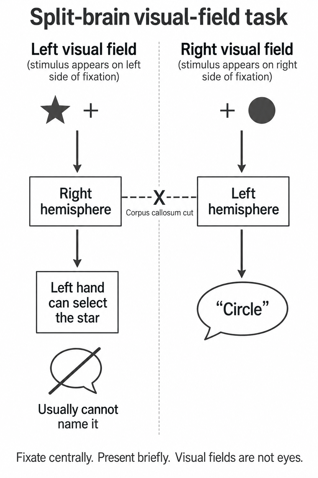
Procedure
Roger Sperry and colleagues, including Michael Gazzaniga, studied split-brain patients using carefully controlled tasks. In a typical visual-field task:
- The participant focuses on a central fixation point.
- A stimulus is flashed very briefly to either the left visual field or the right visual field.
- Because the stimulus is brief, the eyes cannot move to bring it into both visual fields.
- Information from the right visual field goes to the left hemisphere. Each visual field is seen by both eyes: a left visual field is not the same as a left eye.
- Information from the left visual field goes to the right hemisphere.
- If the corpus callosum is cut, the hemisphere that receives the information cannot easily pass it to the other hemisphere.
Continue this lesson.
You’re previewing NovaLearn without an account. Create a free account to read the rest.
Create free account